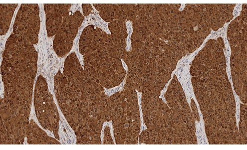
Malignant Melanoma: Immunohistochemistry staining of S-100B scanned using the Aperio AT2. Nuclear and cytoplasmic staining in tumor cells. S-100B: clone EP32
Malignant Melanoma: Immunohistochemistry staining of S-100B scanned using the Aperio AT2. Nuclear and cytoplasmic staining in tumor cells. S-100B: clone EP32
S-100
Antigen Background
S-100A and S-100B proteins are two members of the S-100 family of proteins. S-100A subtypes are composed of one alpha and one beta chain, whereas S-100B is composed of two beta chains. S-100 protein is reported to be expressed in neuroectodermal tissue, including nerves and melanocytes. Langerhans cells in skin are also reported to express S-100 protein. It is noteworthy that S-100 protein is highly soluble and may be eluted from frozen tissue during immunohistochemical procedures.
Product Specific Information
Clone EP32 was raised against S-100 beta protein. Immunoreactivity was observed in neuroectodermal tissue e.g. melanocytes, nerve fibres, dendritic cells, adipocytes and a percentage of macrophages, lymphocytes and plasma cells.
Disclaimer
S-100 is recommended for the detection of specific antigens of interest in normal and neoplastic tissues, as an adjunct to conventional histopathology using non-immunologic histochemical stains.