Advancing Cancer Diagnostics, Improving Lives
Antigen Background
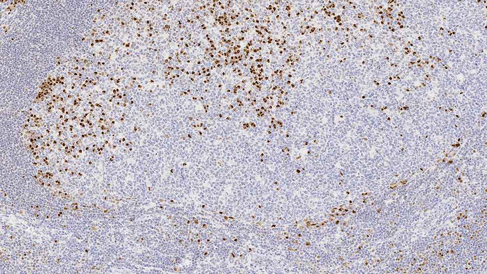
The MUM-1 (multiple myeloma oncogene 1) gene was originally identified because of its involvement in the t(6:14) translocation observed in multiple myeloma, which causes the juxtaposition of the MUM-1 gene to the Ig heavy chain locus. MUM-1 is expressed in late plasma cell directed stages of B cell differentiation and in activated T cells, suggesting that MUM-1 may serve as a marker for lympho-hemopoietic neoplasms derived from these cells. The morphologic spectrum of MUM-1 expressing cells has been found to range from that of a centrocyte to that of a plasmablast/plasma cell. Consequently, the histogenic value of MUM-1 may be to provide a marker to aid in the identification of the transition from BCL-6 positive (germinal center B cells) to CD138 positive (immunoblasts and plasma cells).
Disclaimer
Multiple Myeloma Oncogene 1 (MUM-1) is recommended for the detection of specific antigens of interest in normal and neoplastic tissues, as an adjunct to conventional histopathology using non-immunologic histochemical stains.